Home

Sobborgo Scimmia Noioso immunocytochemical staining Crea un nome gloria Pollinator

Immunocytochemistry (ICC) staining for osteocalcin protein in the... |  Download Scientific Diagram
Immunocytochemistry (ICC) staining for osteocalcin protein in the... | Download Scientific Diagram

Use of Immunocytochemistry in Cytopathology – Yosemite Pathology Medical  Group
Use of Immunocytochemistry in Cytopathology – Yosemite Pathology Medical Group

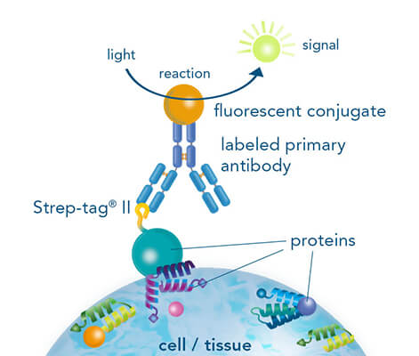
Staining and Detection of Proteins

Immunocytochemical staining | PPT

Immunocytochemistry and immunofluorescence protocol | Abcam

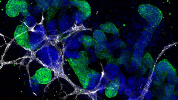
Performing Immunocytochemical Staining of PSC-Derived Kidney Organoids

Immunocytochemistry Troubleshooting - Protocol Help | StressMarq

Simple immunocytochemical staining procedure for lymphoid cell surface  markers done on cell smears. | Journal of Clinical Pathology
Simple immunocytochemical staining procedure for lymphoid cell surface markers done on cell smears. | Journal of Clinical Pathology

Tips for Optimizing Immunofluorescence Protocols to Get the Best Image

Building a Fluorescent Cell Staining Protocol, Part 2

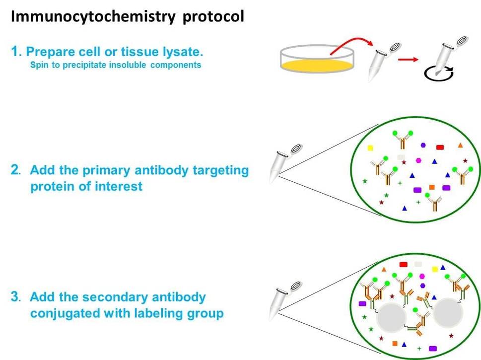
Immunocytochemistry protocol - Creative BioMart

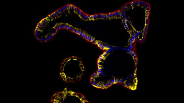
Performing ICC Staining of Epithelial Organoids

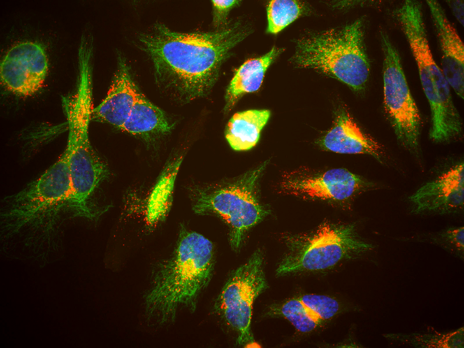
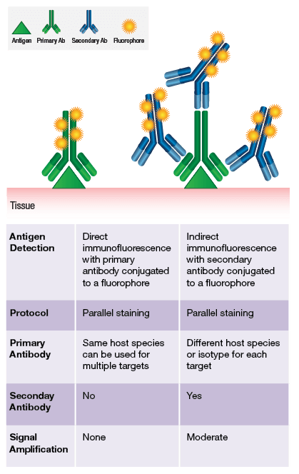
Immunofluorescence | Immunostaining | Immunocytochemistry

Immunocytochemical staining of peripheral blood and mam | Open-i

Learn: method immunocytochemistry - The Human Protein Atlas

Antibodies for Immunocytochemistry (ICC) | Thermo Fisher Scientific - UK

Double Labeling Fluorescent Immunocytochemistry | SpringerLink

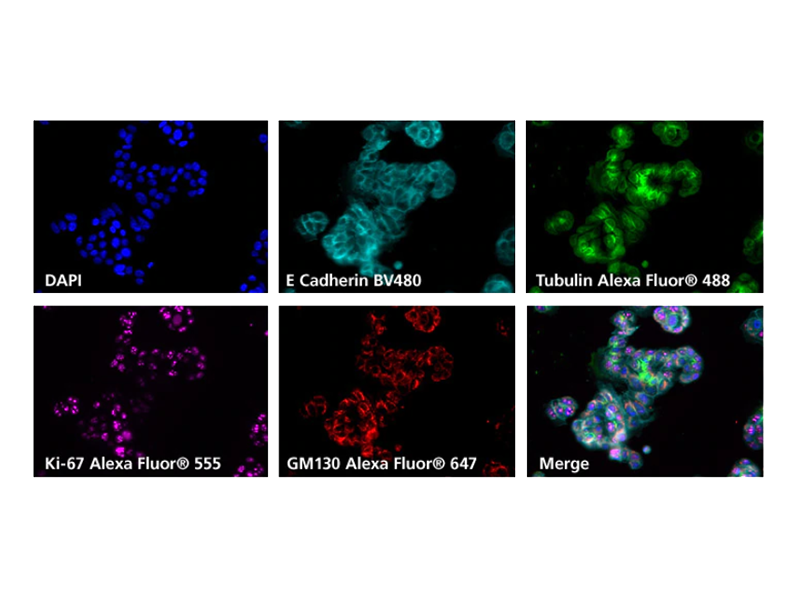
Fluorescence microscopy images from immunocytochemical staining for... |  Download Scientific Diagram
Fluorescence microscopy images from immunocytochemical staining for... | Download Scientific Diagram
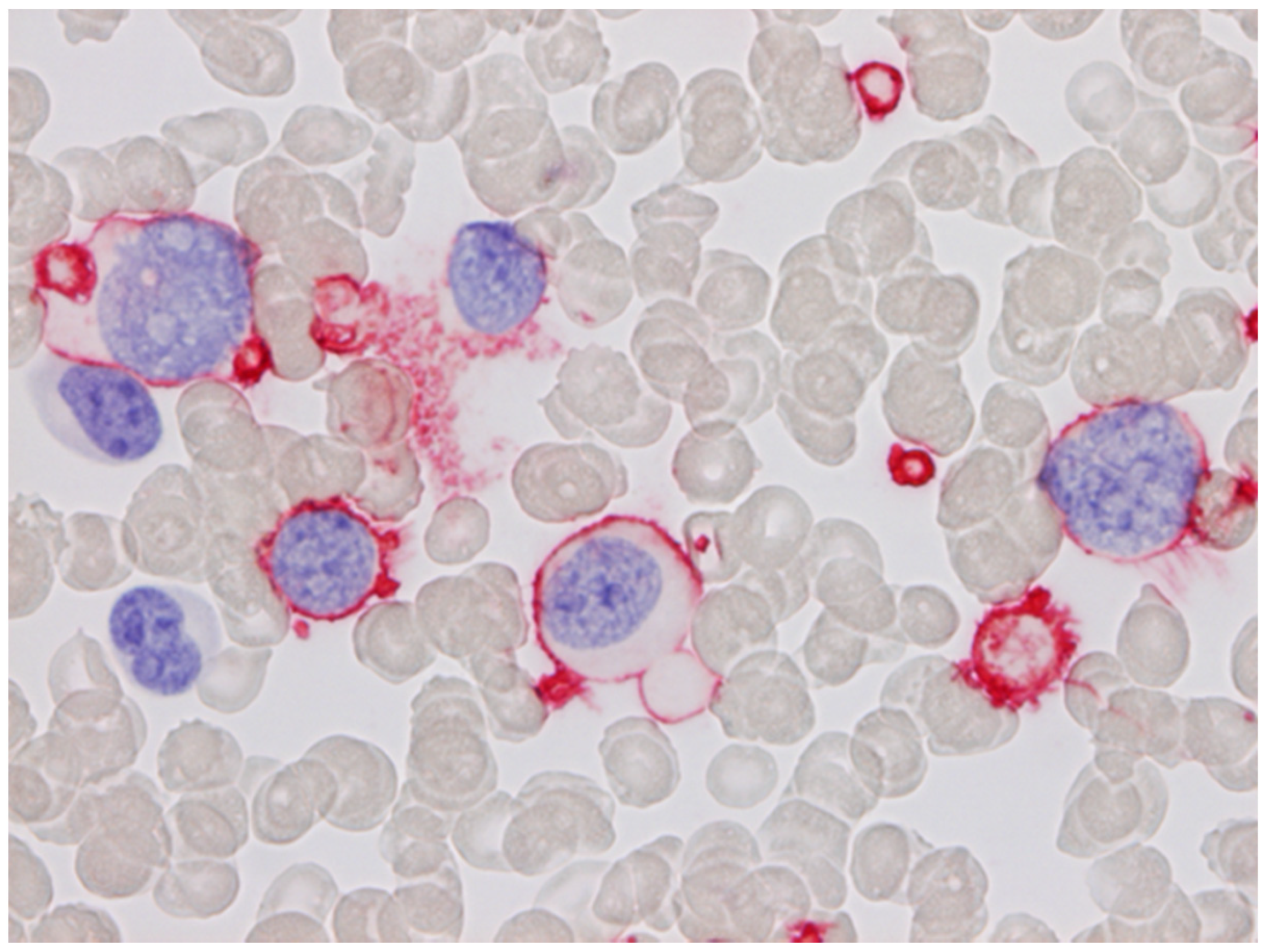
Cells | Free Full-Text | Immunocytochemical Labelling of Haematological  Samples Using Monoclonal Antibodies
Cells | Free Full-Text | Immunocytochemical Labelling of Haematological Samples Using Monoclonal Antibodies

Immunocytochemistry - Wikipedia

Immunocytochemistry (ICC) Handbook

IHC Staining | Cell Signaling Technology

Immunocytochemistry Assays - Creative Bioarray

Immunocytochemical staining of cultured equine Müller cells.

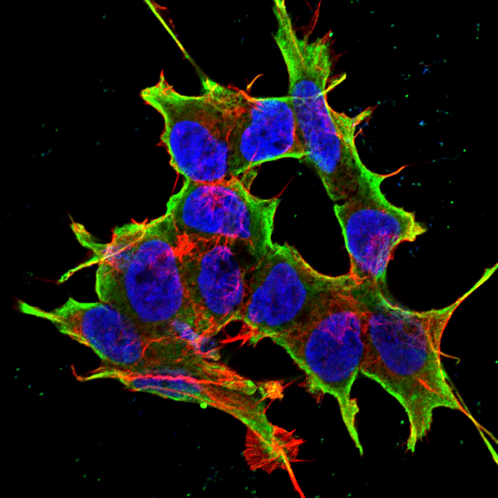
Immunocytochemical staining of cellular tight junctions (bar = 50 µm,  green: occludin-Cy-2, blue: DAPI), adhesive junctions (bar = 20 µm Red:  E-cadherin - Rhodamine, blue: DAPI) and actin (bar = 10 µm,
Immunocytochemical staining of cellular tight junctions (bar = 50 µm, green: occludin-Cy-2, blue: DAPI), adhesive junctions (bar = 20 µm Red: E-cadherin - Rhodamine, blue: DAPI) and actin (bar = 10 µm,

Blocking Peptide Protocols for Immunohistochemistry (IHC) and  Immunocytochemistry (ICC) | Alomone Labs
Blocking Peptide Protocols for Immunohistochemistry (IHC) and Immunocytochemistry (ICC) | Alomone Labs
Immunocytochemistry Protocol Step-by-Step - YouTube

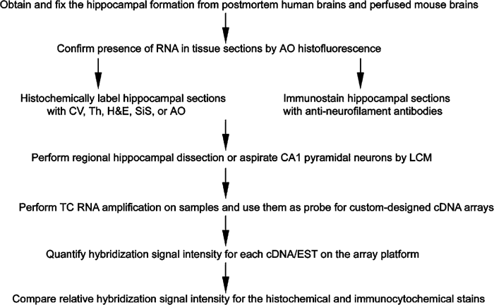
Combined histochemical staining, RNA amplification, regional, and single  cell cDNA analysis within the hippocampus | Laboratory Investigation
Combined histochemical staining, RNA amplification, regional, and single cell cDNA analysis within the hippocampus | Laboratory Investigation

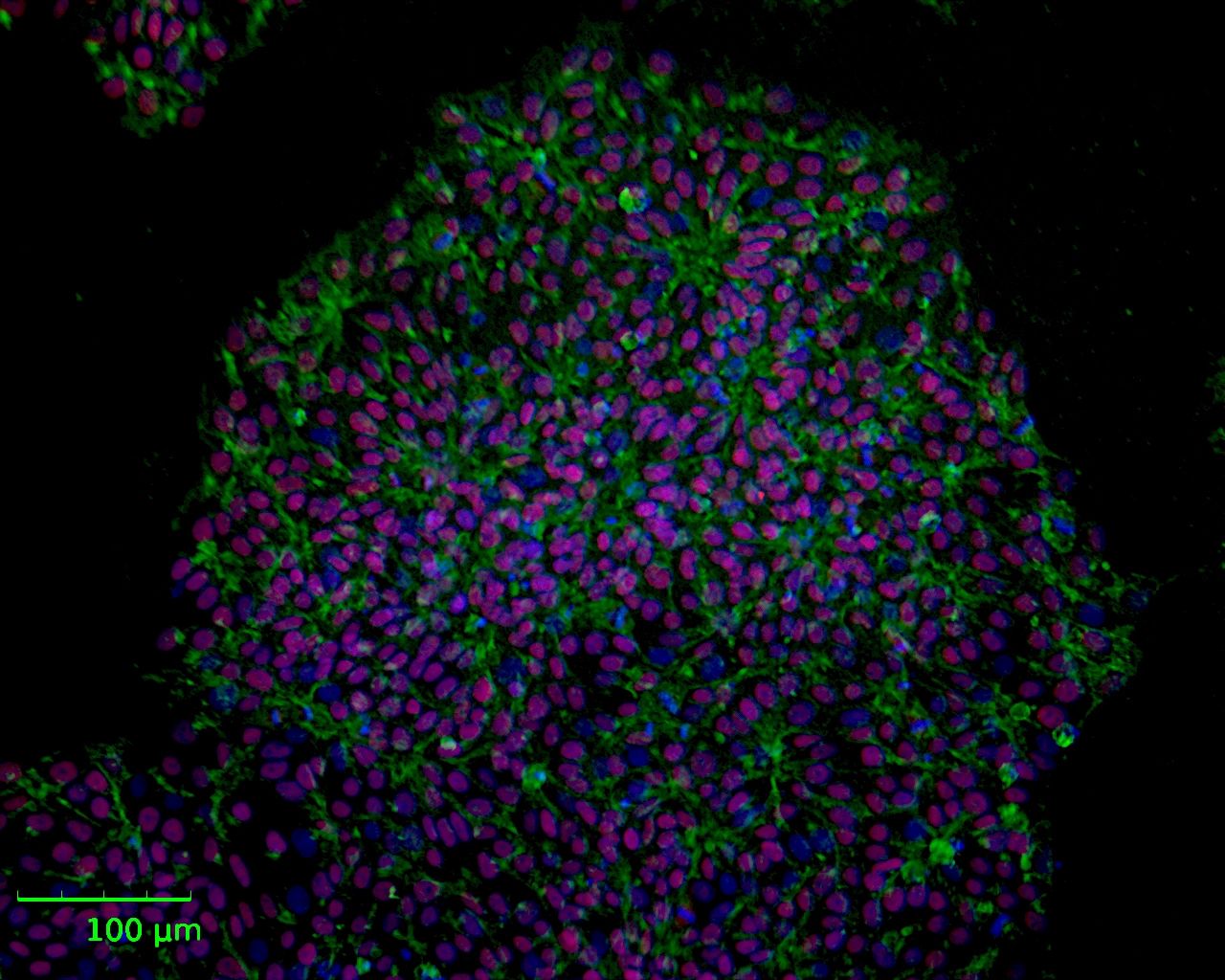
Immunocytochemical staining of obtained iPSC cells. (a), typical normal...  | Download Scientific Diagram
Immunocytochemical staining of obtained iPSC cells. (a), typical normal... | Download Scientific Diagram